
RELATED TOPICS:
EUGLENA:
https://www.sciencearena.in/2025/05/euglena.html
MONOCYSTIS:
https://www.sciencearena.in/2025/05/monocystis.html
PARAMECIUM
STRUCTURE OF PARAMECIUM
1. Size and Shape: Paramecium is unicellular microscopic organism. Its size
varies in different species being 170 µ-290 µ in P. caudatum and 120-250µ in P.
aurelia.
Paramecium (Gr.
Paramaekos or parameces, oblong + L. caudata, tail) is an elongated,
slipper-shaped animal and is commonly referred as slipper animalcule. Its
body is asymmetrical with flat oral or ventral and a convex aboral
or dorsal surface. The anterior end is rounded and the posterior end is thick
and cone-shaped.
The structure is more complicated due to the development of
certain organelles and can be described under the following heads:

2. Pellicle The
body is covered by a thin, firm but elastic pellicle. It gives a definite body form to the organism. The
pellicle is divided into polygonal or hexagonal depressions with raised margins.
A single cilium projects out from the centre of each hexagonal area. The
polygonal areas correspond to regular series of cavities, the alveoli, from
which cilla emerge. The anterior and posterior margins of hexagonal areas bear
the openings of trichocysts.
Pellicle consists of three membranes. The outer or surface membrane is continuous with the membrane surrounding the cilia.
Beneath the outer membrane are closely packed alveoli. These are greatly flattened. The outer and inner membranes
of the alveoli thus form the middle
and inner membranes of the pellicle.
3. Cilia-The
entire body surface is covered by a uniform covering of hair-like protoplasmic
processes, the cilia. These emerge out from the centre of alveoli. All are of
equal size except for a few at the extreme posterior and which are longer and
form the caudal tuft.

Ultrastructure of cilia-Each
cilium consists of fluid matrix surrounded by membranous sheath. The membranous
sheath is continuous with the outer membrane of the pellicle. Within the matrix
are nine peripheral longitudinal fibres
and two central longitudinal fibres.
Each fibre is formed of two subfires, one of which carries a double row of
short arms all running in the same
direction. The central fibres are single and are enclosed within an inner
membranous sheath. Nine very delicate accessory of radial fibrils lie
between the central and peripheral fibres.
1. Cytoplasm
It is distinguished into two regions:
(1) ectoplasm
(2) endoplasm.
Ectoplasm or cortex It is a narrow, peripheral,
dense zone. It includes infraciliary system and trichocysts.
1. Infraciliary system- It consists of basal bodies
(kinetosome) and kinetodesmata located just beneath the alveoli in the ectoplasm.
(a) Basal bodies or kinetosome- The base of each cilium is connected with a tubular basal body or kinetosome.

(b) Kinetodesmata-
From the basal body of each cilium arises a single fibril of kinetosome. It runs anteriorly tapering
along its course. It joins other fibrils of posterior kinetosome of the same
row forming a bundle of fibrils called the kinetodesma.
The individual fibrils do not run anteriorly farther than the five basal
bodies. Thus the number of fibrils in each kinetodesma remains five. The
kinetodesma and the row of kinetosomes associated with form a structural unit
called a kinety. All the kineties
together form infraciliary system. This coordinates beating of
cilia. Infraction plays an important role in the morphogenesis of Paramecium
and other ciliates.
The basal granules are also said to be connected by some
other types of fibrils, the myonemes
and neuronemes. These are highly
contractile and co-ordinate the movement of cilia. All the myonemes converge to
form a darkly stained bilobed body, the motorium,
situated near the cytopharynx. The basal granules, myonemes and motorium
constitute the neuromotor system. It is said to provide a
conducing and coordinating mechanism like the nervous system of higher
animals. But electron microscopy does not reveal the presence of any neuromotor
system.
2. Trichocysts-
These are rod-like or spindle-shaped. These lie embedded in the ectoplasm
alternating with basal bodies and perpendicular to the body surface. Each
trichocyst consists of an elongated shaft
and a terminal spike or barb covered with a cap. The shaft is filled with a dense fluid having a swelling
substance with a fibrous protein.
3. Nuclear apparatus- it consists of a large
bean-shaped macro or meganucleus situated near the cytostome and a small
rounded micronucleus lodged in the depression of meganucleus.
Endoplasm or
medulla- It is the large, central granular, semi-fluid zone. It contains mitochondria, ribosomes, Golgi bodies,
reserve food granules, etc.
4. Contractile apparatus- These are two large
contractile vacuoles, one on either end of the body. Their position is fixed
and they lie between the ectoplasm and endoplasm close to dorsal surface.
5. Food vacuoles- These are roughly spherical,
noncontractile bodies varying in size and number lying in the endoplasm. These
contain ingested food particles.
6. Cytopyge or Cytoproct-It lies on the ventral side
of the body a little behind the cytosome or mouth. It is visible only when the
undigested food particles are eliminated through it.
Nutrition
Paramecium exhibits holozoic
or animal-like mode of nutrition. The process consists of the following steps:
1. Food- Food of
Paramecium consists of bacteria, algae, diatoms, yeasts and other small
protozoans.
2. Ingestion of food-
The food is ingested through cytostome. A current of water is produced by the
constant lashying movement of cilia of oral groove, by which the food particles
are swept towards the cytostome and are claimed down into the cytopharynx.
These are concentrated into a ball by the movement of penniculus and quadrulus.
The ball is finally nipped off from the end of cytopharynx as a food vacuole.

3. Digestion and assimilation- The food vacuoles thus formed are swept by the
streaming movement of endoplasm into the body and are carried along a definite
course. This rotatory streaming movement is known as cyclosis. The food
vacuoles starting from cytopharynx are carried firstly behind, then forward to
the dorsal or aboral surface and finally backward down to the oral surface. The
food is digested inside the food vacuole during its journey. The food vacuole
is first acidic and alkaline later on. The proteins, carbohydrates and fats are
digested. The digested food is assimilated by the endoplasm during cyclosis.
4. Cyclosis: Food
vacuoles are circulated in the body by the streaming endoplasm along a definite
path. This streaming movement is called cyclosis.
Several food vacuoles may be seen circulating in the endoplasm under
microscope. The path followed by food vacuoles is:
From cytopharynx → Down to posterior end→ Upward to become
dorsal → Anterior part→ Backward→ Cytopyge
5. Egestion- The
undigested faecal matter is discharged outside through a definite anal spot or cytopyge
situated posterior to the cytostome.
Locomotion in Paramecium
Paramecium progresses by the following two methods:
1. Ciliary movement- Cilia are main locomotory
organelles in Paramecium. These are fine, hair-like protoplasmic processes all
over the body. These are inclined backward and their beating drives the body
forward. But they may be directed forward and then their strokes push the body
backward. The cilia of a longitudinal row beat one after the other in a metachronial succession or in a metachronous
rhythm. The cilia of transverse row
vibrate simultaneously i.e. synchronously.
The movement of cilia is controlled by the neuromotor system.

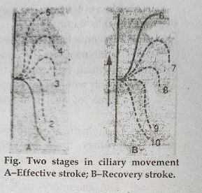
Each oscillation of cilia consists of two strokes:
(i) the effective
stroke in which cilia become
slightly curved and rigid to strike the water like an ore
(ii) recovery stroke in which cilia remain fixed to
offer least resistance to the current.
The cilia beat somewhat towards the right side. As result
the body of Paramecium rotates spirally slightly towards the left. Secondly,
the cilia of oral groove strike more vigorously and obliquely.

2. Body contortions- Paramecium can pass
through a passage narrower than its body by the contraction and twisting of the
body. After this, the body assumes the normal form.
Conjugation in Paramecium
Conjugation is
temporary pairing of two individuals of the same species but from different
mating types for the exchange of their nuclear material. It occurs after
repeated binary fission and is essential for rejuvenation and continuity of
race.
Paramecia ready to pair are sticky and smaller in size. The
individual of two different strains pair with their oral surfaces together. The
pellicle and ectoplasm in the region of union degenerate and a protoplasmic
continuity is established between the two. These are called conjugants. The following nuclear
changes occur in each conjugant simultaneously:
(A) Macro-nuclear changes-
Soon after pairing the macro-nucleus degenerates into fragments and is absorbed
in the cytoplasm.
(B) Micronuclear
behaviour:
(a) Simultaneously, the micronucleus undergoes two pregametic
divisions, of which first is reduction.
As a result four daughter micronuclei are formed each with haploid number of
chromosomes.
(b)Three of the four daughter micronuclei degenerate in each
conjugant.
(c) The remaining micronucleus divides unequally producing a
small active migratory male pronucleus and large and massive stationary pronucleus
potentially female. These are comparable to the nuclei of gametes of high
animals.
(d) The migratory male pronucleus of the two conjugants are
exchanged so that the male pronucleus of one passes into the other and fuses with
the female pronucleus forming the zygote
nucleus or synkaryon.
(e) The conjugants now separate and are called exconjugants.
(f) The synkaryon in each conjugate divides thrice and eight
nuclei are formed. Four of them enlarge and form macronuclei, while the remaining four are known as micronuclei.
(g) Three of the four micronuclei disintegrate.
(h) The single micronucleus in each exconjugant divides
twice and each division is accompanied with the division of body. As a result
four daughter paramecia are formed from each exconjugant each with one micro
and one macronucleus.
(C) Significance of conjugation-
Conjugation is an important process of nuclear reorganisation and nuclear
exchange recurring in between the asexual reproduction. To some extent, it
resembles the sexual reproduction of higher animals, the end of products of
this process are eight daughter paramecia formed after the asexual
multiplication of the exconjugants. The sexual process ends with the fusion of
two gametic nucleic. Therefore, the process is mainly concerned with the
exchange of nucleic material and formation of new macronucleus. It is,
therefore, treated as an episode in reproduction which leads to the following
important events:
1. Rejuvenation-It
has been found that individuals cannot continue to multiply indefinitely by
asexual methods. After a definite number of asexual generations the rate of
fission declines. The individual starts losing its vigour and physiological
efficiency, gives an unhealthy appearance and ultimately dies. The conjugation
is the only prospective measure to regain the former vigour, to revive its
healthy organisation, and thus avoids the senile decay of race.

2. Nuclear reorganisation-The macronucleus is
formed of trophochromatin which governs all the physiological activities of the
individual. Due to repeated fissions its potentiality to coordinate the life
processes ceases and the individual enters a state of depressed physiological
activities. This old and decaying macronucleus is replaced by a new one during
conjugation. This brings about a renewed vigour and vitality.
3. Heredity variations-The fusion of pronucleus
facilitates exchange of nuclear material. This ensures the new combinations of
heritable characters from two different individuals and better adaptability to
the new conditions of life.
Factors Responsible
for Conjugation
1. It does not occur under favourable conditions. Shortage
of food and a particular bacterial diet or certain chemicals induce
conjugation.
2. A certain range of temperature and light, different for
different species is essential for conjugation.
3. The conjugating individuals are usually smaller in size
than the normal individuals.
4. A definite rate of nutrition is necessary for
conjugation.
5. A definite number of binary fissions is necessary for
paramecium to conjugate.
6. Pairing conjugants are isogamous and there is no
morphological sexual dimorphism into male and female conjugants.
7. Conjugation takes place between individuals of different
strains and mating types.
8. Agglutination favours conjugation. It is the interaction
of mating type substances localised in cilia.
Endomixis
Endomixis is
nuclear reorganisation followed by the division of body within a single
individual, with no nuclear exchange or fusion. It occurs only in the absence
of conjugation and has been described by Woodruff
and Erdmann in a binucleate species,
P. aurelia. The process can be distinguished into the following steps:

1. The macronucleus disintegrates and is absorbed in the
surrounding cytoplasm.
2. The two micronuclei divide twice forming eight daughter
micronuclei.
3. Six of them disappear leaving two micronuclei.
4. The animal with two micronuclei divides by transverse
binary fission.
5. Micronucleus in each daughter Paramecium divides twice.
6. Of these four nuclei, two enlarge to form two macronuclei
and two micronuclei.
7. The micronuclei in each daughter individual again divide
accompanied by the division of body.
Thus four daughter paramecia are formed from a single parent
and each possesses one macronucleus and two micronuclei.
Significance of Endomixis
1. Multiplication:
As a result of endomixis four individuals are formed from a single parent.
2. Rejuvenation:
The effect of endomixis is similar to that of conjugation, because the old
macronucleus is replaced by a new one formed from the micronuclear material.
This leads to renewed vigour and vitality.
3. The endomixis is regarded as a substitute for conjugation
since it occurs only when conjugation is prevented or delayed.
RELATED TOPICS:
EUGLENA:
https://www.sciencearena.in/2025/05/euglena.html
MONOCYSTIS:
https://www.sciencearena.in/2025/05/monocystis.html
FOLLOW
THE INSTRUCTIONS FOR DOWNLOAD THIS ASSIGNMENT:
1. SEARCH WWW.SCIENCEARENA.IN
2. OPEN EDUCATION AND RESOURCES IN MENU BAR.
3. SINGLE CLICK DOWNLOAD AVAILABLE IN DOWNLAOD SECTION.
https://www.sciencearena.in/p/education-resources.html